
-
Research Services<a class="dhimg1" href="/shichangdetail1/9.html" target="_self" rel="nofollow"> <img src="https://omo-oss-image.thefastimg.com/portal-saas/pg2026042316544414203/cms/image/8926ff01-64bc-4928-a995-396bdb9d8102.jpg" alt="Aniphe BioLab"> </a> <a class="dhimg2" href="/shichangdetail/8.html" target="_self" rel="nofollow"> <img src="https://omo-oss-image.thefastimg.com/portal-saas/pg2026042316544414203/cms/image/26342b34-4f1e-4d28-9166-7b685e6d70c3.jpg" alt="Aniphe BioLab"> </a>


-
Preclinical CRO<a class="dhimg1" href="/shichangdetail/6.html" target="_self" rel="nofollow"> <img src="https://omo-oss-image.thefastimg.com/portal-saas/pg2026042316544414203/cms/image/cc43bac1-07d9-4340-9677-fbe01c51330a.jpg" alt="Aniphe BioLab"> </a> <a class="dhimg2" href="/shichangdetail/10.html" rel="nofollow" target="_self"> <img src="https://omo-oss-image.thefastimg.com/portal-saas/pg2026042316544414203/cms/image/f1d8f739-0d36-4cb8-adeb-26de791d909c.jpg" alt="Aniphe BioLab"> </a>


-
-
-
-


Latest News
Focusing on cutting-edge technologies and research advancements,
sharing Aniphe BioLab’s achievements in scientific innovation,
Professional insights
and practical experience in the fields of laboratory animals and preclinical research.
Publish an article
Key Insights | APP/PS1 Double-Transgenic Mice: Cognitive Function, Hippocampal Aβ, and Telomere Length—Everything You Want to Know Is Right Here!
Introduction:
APP/PS1 is a double-transgenic mouse model that replicates key pathological features of Alzheimer’s disease, including amyloid plaque formation and cognitive decline, and is widely used in AD research. This article examines the model from multiple perspectives, including body weight, cognitive function, hippocampal Aβ levels, and telomere length (TL).
——————————————————
As China gradually transitions into an aging society, the number of individuals with Alzheimer’s disease (AD) is projected to reach 16.46 million by 2030, accounting for approximately one-quarter of the global total. This will impose a substantial medical and economic burden on both families and society.
The pathological features of Alzheimer’s disease (AD) are complex, and its underlying mechanisms remain poorly understood. Currently, clinical AD‑targeted therapies can only slow the progression of symptoms; they neither effectively halt nor reverse the disease course, and relapse often occurs upon discontinuation of treatment. Consequently, the development and establishment of animal models for this condition are of paramount importance. Today, we introduce one of the most widely used AD animal models: the APP/PS1 mouse.
APP/PS1 double-transgenic mice
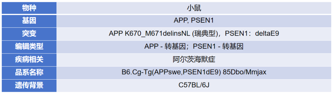
Image source: Aniphe BioLab
The APP/PS1 mouse is a double-transgenic model that expresses a chimeric murine/human amyloid precursor protein (Mo/HuAPP695swe) and a mutant human presenilin 1 (PS1-dE9). It mimics key pathological features of Alzheimer’s disease, including amyloid plaque formation and cognitive decline, and is widely used in AD research.
The CNS comprises billions of neurons and serves as the structural basis for interneuronal communication. Both Mo/HuAPP695swe and PS1-dE9 are targeted to CNS neurons. The “humanized” Mo/HuAPP695swe transgene enables mice to secrete human A‑β peptides. Both the transgenic peptides and the full-length protein can be detected by antibodies specific to the corresponding human sequences in that region. The transgenic mutant human presenilin protein (PS1-dE9) replaces detectable endogenous mouse protein at high levels and is also immunodetectable in whole-brain protein lysates.
During the aging process, the brains of APP/PS1 mice develop amyloid plaques and exhibit declines in cognitive and behavioral functions, along with other AD‑related pathological changes.

Its main features include the development of abnormal amyloid plaques in the brains of APP/PS1 mice during aging, cognitive impairment characterized by declines in learning and memory, and observable neuronal degeneration and synaptic damage, including reduced synaptic density and neuronal loss.
Body weight differences in mice as they age
Studies have found that, under conditions of identical genotype and age, with the exception of 8-month-old APP/PS1 double-transgenic mice, female mice of all other strains weigh less than their male counterparts.
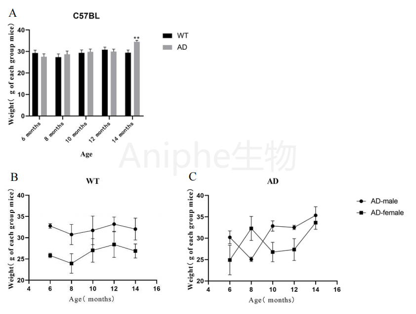
The effects of age and sex on the body weight of AD mice; image source [3]
In comparisons among mice of the same age but different genotypes, with the exception of 14-month-old APP/PS1 double-transgenic mice, the body weights of the other mice remained relatively stable across age, without significant differences. The largest weight difference observed among the APP/PS1 double-transgenic mice was approximately 7 g, and all mice weighed between 20 g and 40 g.
The magnitude of cognitive decline in APP/PS1 double-transgenic mice tends to diminish with increasing age, and aging itself contributes to the deterioration of cognitive function.
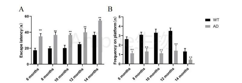
Morris water maze spatial exploration experiment; image source [3]
Compared with 6-month-old mice, 14-month-old APP/PS1 double-transgenic mice showed an increase of approximately 20 seconds in escape latency. At 6 months of age, the escape latency of these mice was up to 19 seconds longer than that of wild-type mice, and the number of platform crossings decreased significantly with increasing age.
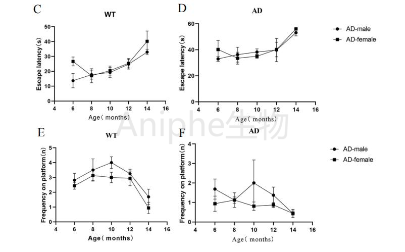
Sex differences in spatial exploration in the water maze task; image source [3]
Under identical phenotypic conditions, comparison between male and female mice revealed that APP/PS1 double-transgenic males exhibited a slightly higher number of platform crossings than females. Increasing age also reduced both the escape latency and the number of platform crossings in both sexes, though no significant differences were observed between males and females.
Aβ production in the mouse hippocampus increases with age.
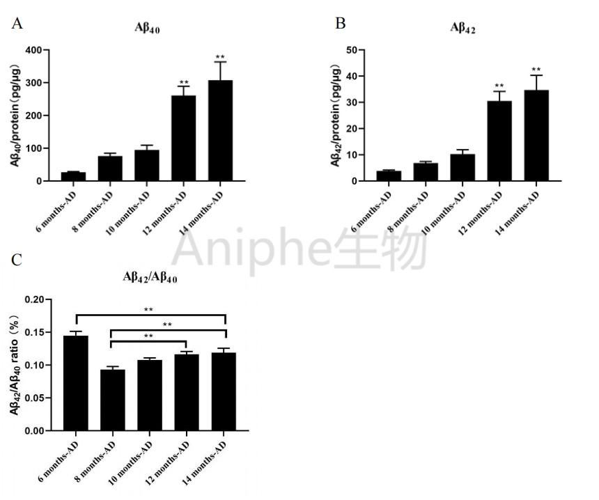
The effect of chronological age on Aβ production in the hippocampus of the AD group; image source [3].
ELISA assay results indicate that, with increasing age, the levels of Aβ40 and Aβ42 in the hippocampus of APP/PS1 double-transgenic mice progressively rise, showing a highly significant increase between 6 and 12 months of age.
At 6 months of age, the APP/PS1 group exhibited relatively low levels of Aβ40 and Aβ42 deposition compared with other ages; consequently, the Aβ42/Aβ40 ratio was elevated relative to the other age groups. Starting at 8 months of age, this ratio continued to increase with advancing age, and by 14 months, it had risen by 4% compared with the 8-month‑old APP/PS1 double-transgenic mice.
With advancing age, Aβ40 and Aβ42 deposition in the hippocampus of APP/PS1 double-transgenic mice increases, and the Aβ42/Aβ40 ratio gradually rises. Compared with 6-month-old mice, the differences in hippocampal Aβ40, Aβ42, and the Aβ42/Aβ40 ratio at 14 months of age reach statistical significance (P = 0.003).
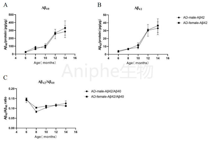
As chronological age increases, sex influences Aβ production in the hippocampus of the AD group; image source [3].
In APP/PS1 double-transgenic female and male mice, hippocampal levels of Aβ40, Aβ42, and the Aβ42/Aβ40 ratio all increased with age. Comparison between female and male APP/PS1 double-transgenic mice revealed no significant sex‑dependent differences, with similar concentrations of Aβ40 and Aβ42 and comparable Aβ42/Aβ40 ratios.
Mouse TL shortens with increasing age.
Experimental measurements of cellular telomere length (TL) were conducted in six organs or tissues of mice, including the prefrontal cortex, pituitary gland, hippocampus, and cerebellum in neural tissue; abdominal skin in epithelial tissue; and the colon, which is composed of multiple tissues. The TL assay results revealed that, across all six organs or tissues, cellular TL in APP/PS1 double-transgenic mice progressively shortened with increasing age.
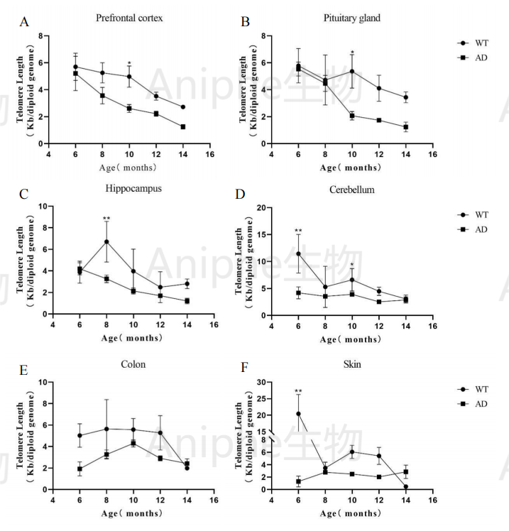
The effect of increasing age on telomere length in AD mice; image source [3]
Under different sex conditions, the effects of increasing lunar age on telomere length (TL) in various tissues of AD mice revealed that, under the same phenotypic conditions, female mice exhibited shorter telomeres in four brain regions—prefrontal cortex, pituitary gland, hippocampus, and cerebellum—compared with males.
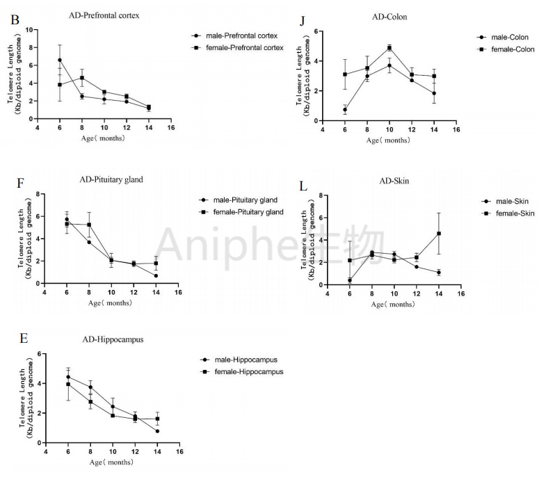
The effects of increasing age and sex differences on telomere length in AD mice; image source: [3]
The incidence of MNPCE in mice increases with advancing age.
When assessing the overall level of genetic damage in AD mice using the MNPCE assay, it was observed that, in both the WT and AD groups, the frequency of micronucleated polychromatic erythrocytes (MNPCE) in bone marrow increased with advancing age.
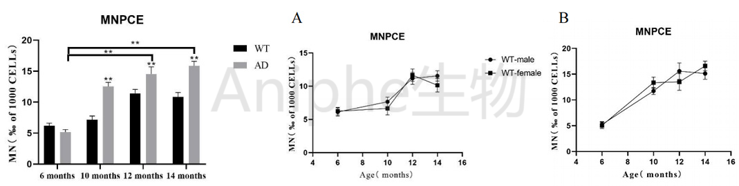
The effect of increasing postnatal age on MNPCE in AD mice and sex differences; image source [3]
At 10, 12, and 14 months of age, MNPCE levels showed a highly significant increase compared with those at 6 months. Under identical phenotypic conditions, no significant differences were observed in MNPCE between male and female mice of the same age; the incidence of MNPCE was similar across both sexes.
As mice age, Increased plaque deposition, Gliosis
APP/PS1–mediated microglial activation precedes amyloid plaque deposition, with microglial proliferation evident by 3 months of age and further augmented at 4 and 6 months. Amyloid plaques in the cerebral cortex begin to emerge around 4 months of age, while those in the hippocampus appear approximately at 6 months, and both continue to accumulate with advancing age.
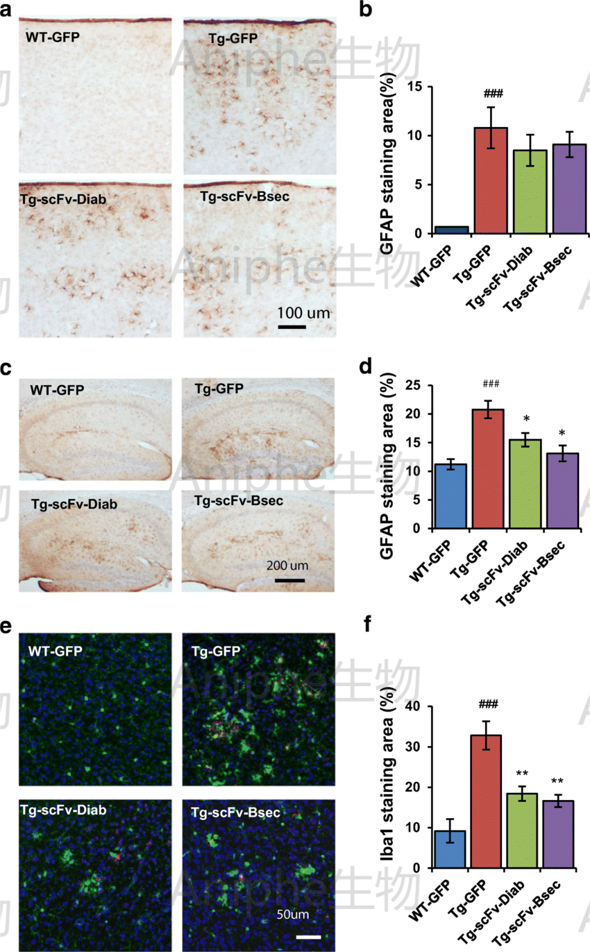
Amyloid‑β deposits in brain sections of APP/PS1 mice; image source: [1].
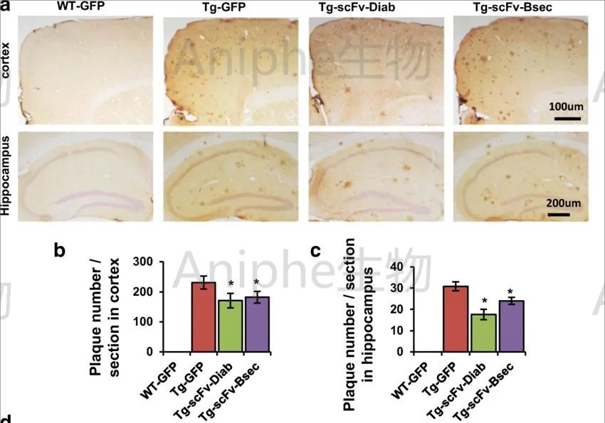
Astrogliosis responding to Aβ in the brains of APP/PS1 mice; image source: [1]
Neuronal loss begins to appear in mice starting in August.
In the brains of APP/PS1 mice, alterations involving neuronal degeneration and synaptic damage can also be observed, including a reduction in synaptic density and neuronal loss.
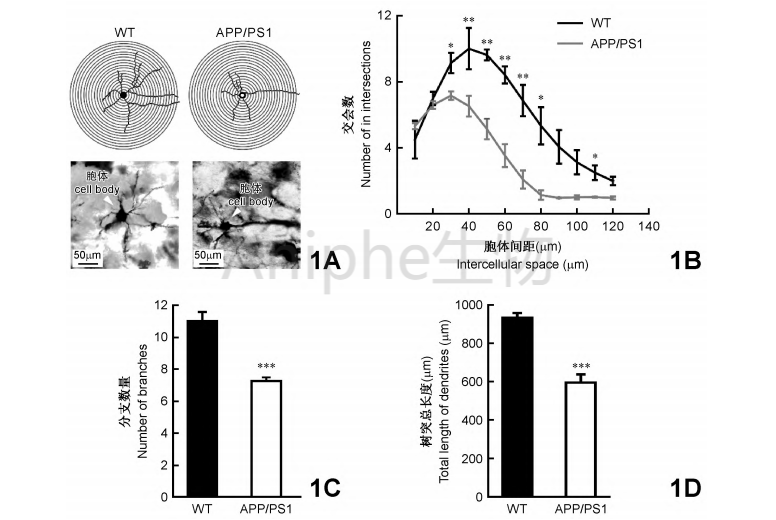
Comparison of the morphology of OB neurons in APP/PS1 and WT mice; image source [5].
Application areas
Mechanism of amyloid plaque formation: APP/PS1 mice can be used to study the formation of amyloid plaques, the metabolism and aggregation mechanisms of β‑amyloid protein, as well as intervention strategies involving relevant therapeutic agents.
Research on Cognitive and Behavioral Functions: Behavioral assays can be used to assess changes in learning, memory, and cognitive function in APP/PS1 mice, thereby facilitating investigations into the pathogenic mechanisms of Alzheimer’s disease and the development of potential therapeutic strategies.
Research on Neurodegeneration and Synaptic Damage: APP/PS1 mice can be used to investigate the mechanisms underlying neuronal degeneration and synaptic dysfunction in Alzheimer’s disease, as well as to evaluate neuroprotective strategies.
Precautions for Mouse Husbandry
Key points for rearing and breeding:
1. APP/PS1 mice are prone to aggression; during housing, they should be closely monitored. If fighting occurs among cage mates, the aggressive individuals should be separated. In cases of severe aggression resulting in bite wounds, treat the injuries with povidone‑iodine or an antibiotic ointment.
2. Disinfectable toys can be placed inside the cage to reduce the likelihood of mice fighting.
3. In the C57BL/6 background, APP mice exhibit a mortality rate of 10–15%.
————————————
References:
[1]Chintapaludi SR, Uyar A, Jackson HM, et al. Staging Alzheimer's Disease in the Brain and Retina of B6.APP/PS1 Mice by Transcriptional Profiling. Journal of Alzheimer's Disease : JAD. 2020 ;73(4):1421-1434. DOI: 10.3233/jad-190793. PMID: 31929156; PMCID: PMC9066610.
[2]Kater MSJ, Huffels CFM, Oshima T, Renckens NS, Middeldorp J, Boddeke EWGM, Smit AB, Eggen BJL, Hol EM, Verheijen MHG. Prevention of microgliosis halts early memory loss in a mouse model of Alzheimer's disease. Brain Behav Immun. 2023 Jan;107:225-241. doi: 10.1016/j.bbi.2022.10.009. Epub 2022 Oct 18. PMID: 36270437.
[3] Li Jianfei. Analysis of Genetic Damage Characteristics in the Nervous System and Other Organs of APP/PS1 Mice [D]. Yunnan Normal University, 2022. DOI: 10.27459/d.cnki.gynfc.2022.001253.
[4]034832 - APP/PS1 Strain Details (jax.org).
[5] Chen Boyu, Jiang Jinxiang, Zhang Jiawei, et al. Abnormalities in early dendritic branching and length of olfactory bulb neurons, as well as in the expression of potassium–chloride cotransporter 2 protein, in APP/PS1 mice [J]. Acta Anatomica Sinica, 2023, 54(02):127–133. DOI: 10.16098/j.issn.0529-1356.2023.02.001.
[6]He, Ping & Xin, Wei & Sierks, Michael. (2019). Bispecific Antibody Fragment Targeting APP and Inducing α-Site Cleavage Restores Neuronal Health in an Alzheimer’s Mouse Model. Molecular Neurobiology. 56. 10.1007/s12035-019-1597-z.
Aniphe BioLab Inc.
Address:
Runcheng, No. 8 Longtai Road, Jiangbei New Area, Nanjing City, Jiangsu Province
Building 8, Science Park
Postal Code: 210007
Phone:025-8520 8061
Email:info@aniphebio.com(Consultation)
Email:order@aniphebio.com(Order)


WeChat Official Account
Video Account
 Su Public Security Bureau Internet Information Service Filing No. 32010402001421