
-
Research Services<a class="dhimg1" href="/shichangdetail1/9.html" target="_self" rel="nofollow"> <img src="https://omo-oss-image.thefastimg.com/portal-saas/pg2026042316544414203/cms/image/8926ff01-64bc-4928-a995-396bdb9d8102.jpg" alt="Aniphe BioLab"> </a> <a class="dhimg2" href="/shichangdetail/8.html" target="_self" rel="nofollow"> <img src="https://omo-oss-image.thefastimg.com/portal-saas/pg2026042316544414203/cms/image/26342b34-4f1e-4d28-9166-7b685e6d70c3.jpg" alt="Aniphe BioLab"> </a>


-
Preclinical CRO<a class="dhimg1" href="/shichangdetail/6.html" target="_self" rel="nofollow"> <img src="https://omo-oss-image.thefastimg.com/portal-saas/pg2026042316544414203/cms/image/cc43bac1-07d9-4340-9677-fbe01c51330a.jpg" alt="Aniphe BioLab"> </a> <a class="dhimg2" href="/shichangdetail/10.html" rel="nofollow" target="_self"> <img src="https://omo-oss-image.thefastimg.com/portal-saas/pg2026042316544414203/cms/image/f1d8f739-0d36-4cb8-adeb-26de791d909c.jpg" alt="Aniphe BioLab"> </a>


-
-
-
-


Latest News
Focusing on cutting-edge technologies and research advancements,
sharing Aniphe BioLab’s achievements in scientific innovation,
Professional insights
and practical experience in the fields of laboratory animals and preclinical research.
Publish an article
Disease Models | Construction of Tumor Animal Models: Osteosarcoma Edition
Osteosarcoma (OS) It is the most common primary malignant bone tumor, predominantly affecting adolescents, characterized by high malignancy, aggressive local invasion, and early pulmonary metastasis, thereby posing a serious threat to patients’ health and survival. Establishing an appropriate animal model can facilitate a deeper understanding of the pathogenesis and therapeutic strategies for bone tumors, providing robust support for clinical diagnosis and treatment.
Currently commonly used osteosarcoma animal models include: Xenograft models, inducible osteosarcoma models, and genetically engineered mice.
01 Transplantable model
In the transplantable model approach, by injection of Human or murine osteosarcoma cells are implanted into immunodeficient mice to establish xenograft or allograft models. This research approach is widely employed, with injected cells giving rise to locally growing solid tumors within a few days after implantation.
According to the injection site, it mainly includes Subcutaneous injection (for studying growth alone) and orthotopic injection (for tumor growth and metastasis).
Selection of Mice
Given the inherent variability in tumor‑formation rates and pulmonary metastasis following transplantation in mice, the selection of recipient animals is of critical importance. Typically, neonatal or immunodeficient strains are chosen, such as nude mice, SCID mice, and NOD‑SCID mice.
Modeling method
Below, we take... In situ injection of human osteosarcoma 143B cells model Taking the experimental procedure for tibial injection as an example:
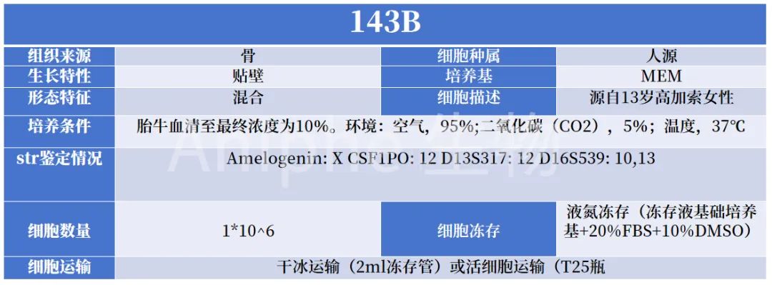
Image source: Aniphe BioLab
01 Recover osteosarcoma cells by culturing them, and collect osteosarcoma cells in the logarithmic growth phase.
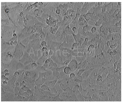
The normal morphology of the 143B osteosarcoma cell line in the logarithmic growth phase; Source [1]
02 Inoculate the cells at the specified concentration into the right axillary region of the mouse’s forelimb.
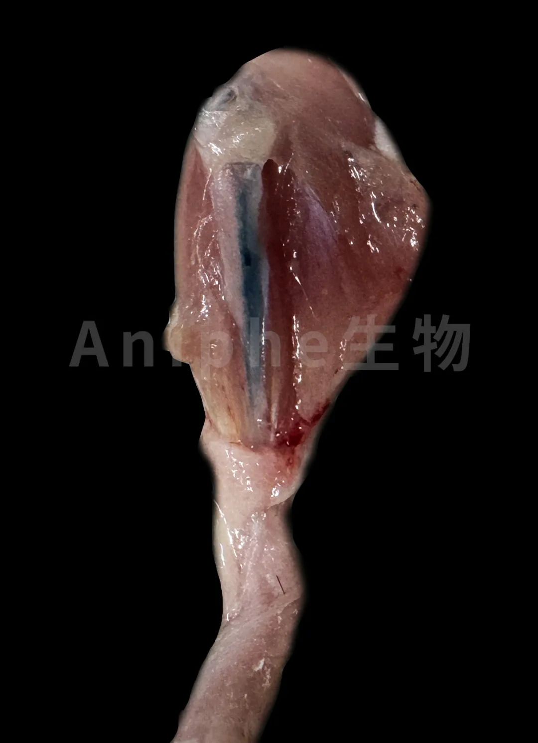
In situ tibial positioning visualization: post‑injection with Evans blue; image source: Aniphe BioLab.
03 Subcutaneous injections were administered, and the tumor’s long and short diameters were measured starting on day 4. Once the tumors had grown to approximately 5 mm in size (around days 10–11 post‑tumor implantation), 68Ga‑DOTA‑NOC PET/CT and 18F‑FDG PET/CT imaging were performed.

Figure 143B shows the tumor growth curve, which gradually increases over time; image source [1].
Model Features
Cells were injected in situ into the tibias of immunodeficient mice, and tumors developed rapidly within 14–21 days after injection, resulting in lytic primary tumors in the mouse tibias.
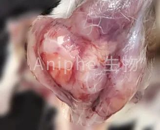
Image source: Aniphe BioLab
micro-CT
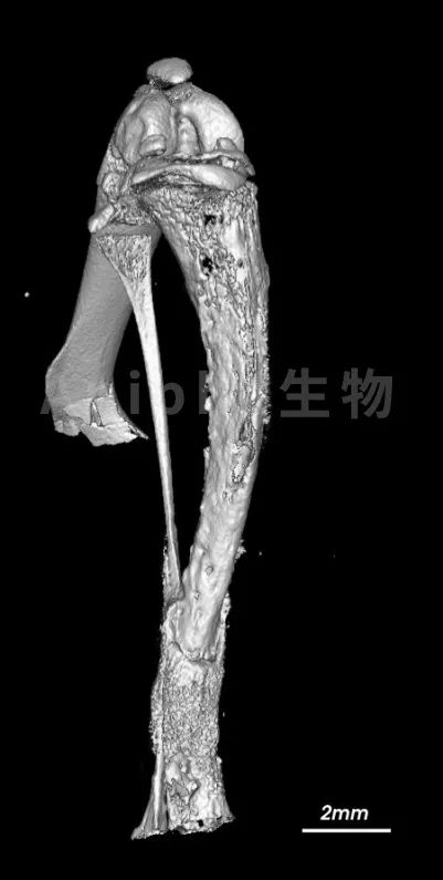
Image source: Aniphe BioLab
As shown in the figure, the mouse bone tissue is destroyed, and irregular soft-tissue masses are observed, accompanied by varying degrees of lamellar periosteal reaction, bone resorption, and subsequent bone damage following periosteal formation.
H&E staining
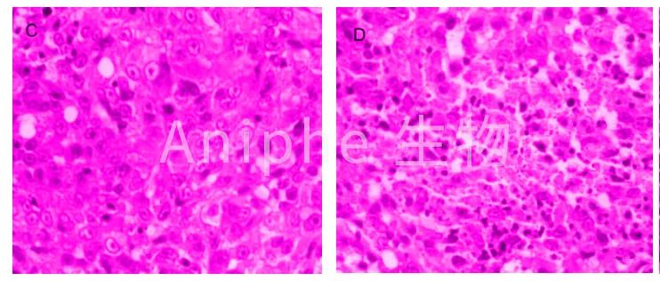
C denotes the tumor morphology; D denotes the morphology of the necrotic portion of the tumor. Image source: [1]
As shown in the figure, the tumor cells exhibit large nuclei with deeply stained chromatin, and necrosis is present within the tumor tissue.
02 Inducible tumor model
An inducible osteosarcoma animal model refers to a system in which physical, chemical, or biological pathogenic factors are applied to animals, resulting in specific damage to tissues, organs, or the entire organism and leading to osteosarcomatous lesions that mimic certain functional, metabolic, or morphological–structural features of human disease—thus artificially inducing osteosarcoma in animals that closely resemble the human condition. For example:
Physical factors: Radionuclides are potent carcinogens, and nearly all bone-seeking radionuclides can induce osteosarcoma.
Chemical factors: Malignant osteosarcomas were induced by continuous intramuscular injection of chemical agents—including zinc beryllium silicate, aflatoxin B1, 4-hydroxyaminoquinoline, 1-oxide of quinoline, beryllium oxide, methylcholanthrene, the copper complex of N-hydroxy-2-acetamidofluorene, and diethylnitrosamine—at specific concentrations.
Virus: Osteosarcoma models are established by injecting viral strains such as Moloney sarcoma virus and SV40 virus.
03 Genetically engineered mice
Genetic knockout and knock-in models in animals enable more accurate simulation of the initiation and progression of osteosarcoma, exhibiting strong concordance with the tumor’s natural developmental trajectory. Moreover, these models can recapitulate certain precancerous lesions, thereby addressing the limitations of xenograft models in mimicking clinical patient scenarios.
Genes such as p53, Rb, p62, p15INK4b, p16INK4a, c-Fos, SV40 T antigen, c-MYC, and VEGF have been found to exhibit abnormalities—including deletions, mutations, or overexpression—in osteosarcoma cases.
At present, the most well-established genetic engineering model—characterized by 100% penetrance—is tissue-specific knockout. p53, Rb Genetically engineered mice and tissue-specific overexpression SV40 T Mouse model of the antigen, NICD (intracellular domain of Notch1).
Osx-Cre+p53 fl/fl pRb fl/fl Mouse In the osteogenic system, Cre expression driven by the tissue-specific promoter Osx enables tissue-specific knockout. p53, pRb Genetically, knockout of pRB promotes the development of osteosarcoma, enabling the generation of mouse models with a 100% incidence of osteosarcoma.
Col1a1 2.3kb-Cre; Rosa26 NICD Mouse Conditional Rosa26NICD mice were crossed with Col1a1 2.3kb-Cre mice to generate the genotyped strain Col1a1 2.3kb-Cre; Rosa26NICD. In this model, the combined activation of Notch signaling and deletion of p53 synergistically accelerate the development of osteosarcoma in mice.
Osteocalcin-SV40 T antigen Mouse Using the osteocalcin promoter ( osteocalcin promoter ) Drive SV40T/t antigen (TAg) expression. Specifically, the large T antigen in TAg targets and inactivates p53 and Rb, while the small T antigen targets PP2A, thereby inducing tumorigenesis. This approach can also achieve a 100% tumor incidence rate.
Aniphe BioLab Inc.
Address:
Runcheng, No. 8 Longtai Road, Jiangbei New Area, Nanjing City, Jiangsu Province
Building 8, Science Park
Postal Code: 210007
Phone:025-8520 8061
Email:info@aniphebio.com(Consultation)
Email:order@aniphebio.com(Order)


WeChat Official Account
Video Account
 Su Public Security Bureau Internet Information Service Filing No. 32010402001421