
-
Research Services<a class="dhimg1" href="/shichangdetail1/9.html" target="_self" rel="nofollow"> <img src="https://omo-oss-image.thefastimg.com/portal-saas/pg2026042316544414203/cms/image/8926ff01-64bc-4928-a995-396bdb9d8102.jpg" alt="Aniphe BioLab"> </a> <a class="dhimg2" href="/shichangdetail/8.html" target="_self" rel="nofollow"> <img src="https://omo-oss-image.thefastimg.com/portal-saas/pg2026042316544414203/cms/image/26342b34-4f1e-4d28-9166-7b685e6d70c3.jpg" alt="Aniphe BioLab"> </a>


-
Preclinical CRO<a class="dhimg1" href="/shichangdetail/6.html" target="_self" rel="nofollow"> <img src="https://omo-oss-image.thefastimg.com/portal-saas/pg2026042316544414203/cms/image/cc43bac1-07d9-4340-9677-fbe01c51330a.jpg" alt="Aniphe BioLab"> </a> <a class="dhimg2" href="/shichangdetail/10.html" rel="nofollow" target="_self"> <img src="https://omo-oss-image.thefastimg.com/portal-saas/pg2026042316544414203/cms/image/f1d8f739-0d36-4cb8-adeb-26de791d909c.jpg" alt="Aniphe BioLab"> </a>


-
-
-
-


Latest News
Focusing on cutting-edge technologies and research advancements,
sharing Aniphe BioLab’s achievements in scientific innovation,
Professional insights
and practical experience in the fields of laboratory animals and preclinical research.
Publish an article
Disease Model | Establishment of a Mouse Myocardial Ischemia Model via Thoracic Compression to Ligate the Left Anterior Descending Coronary Artery (LDA)
Introduction Myocardial ischemia (MI) refers to a pathological condition in which reduced cardiac blood perfusion leads to diminished oxygen supply to the heart, resulting in abnormal myocardial energy metabolism and an inability to sustain normal cardiac function. With the emergence of an aging population in China, cardiovascular and cerebrovascular diseases have increasingly become the leading causes of morbidity and mortality. Establishing an appropriate animal model of myocardial infarction is therefore of paramount importance for investigating the pathogenic mechanisms of MI and developing effective prevention and treatment strategies.
——————————————————————
At present, several methods are available for establishing animal models of myocardial ischemia, including coronary artery ligation, pharmacological induction, balloon occlusion, embolization, and thrombosis induction. Among these, Coronary artery ligation method The procedure is simple, with clear visualization of vascular occlusion, closely mirroring the pathophysiological process of myocardial infarction and facilitating effective clinical translation.
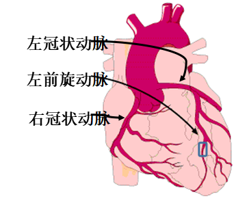
And Chest compression method for ligating the left anterior descending coronary artery (LAD) A mouse model of myocardial ischemia is established by creating a small incision to expose the heart, without the need for mechanical ventilation. This approach is widely adopted due to its rapidity, minimal invasiveness, and low perioperative mortality.
Method for establishing a myocardial infarction model in mice
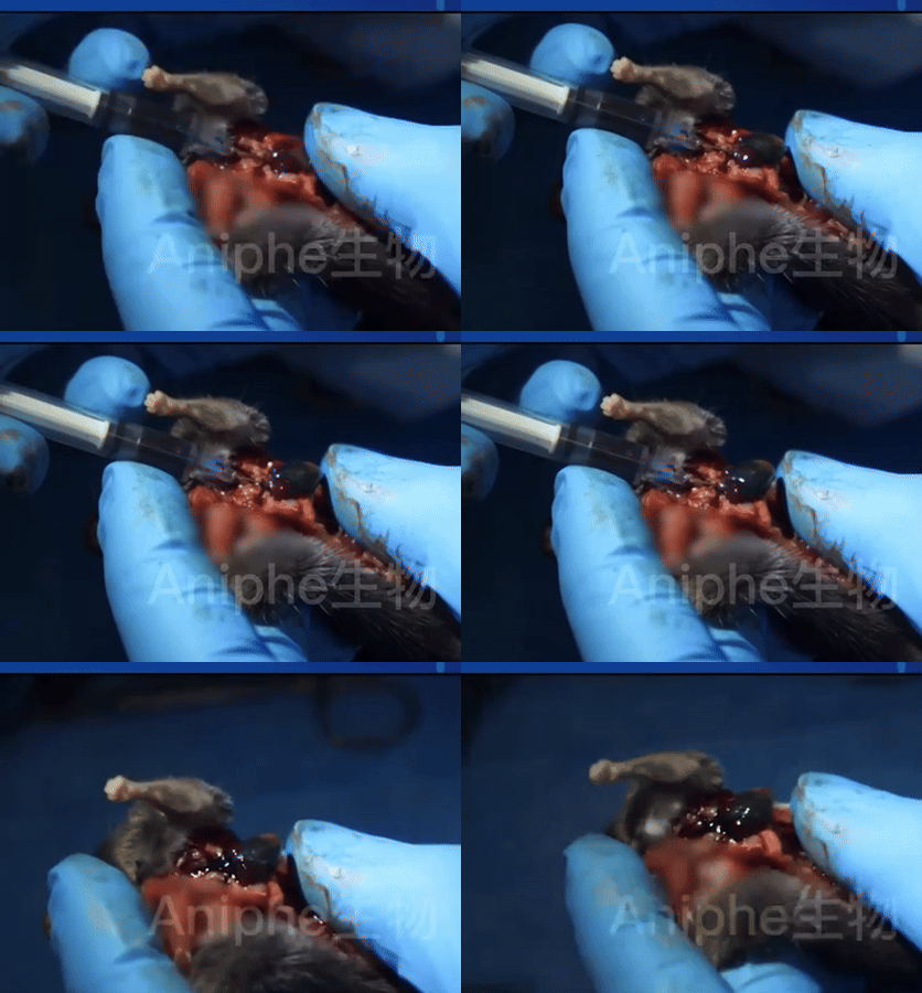
01 Mice were anesthetized and immobilized in the supine position;
02 A longitudinal incision is made at the skin over the left sternal border, and a vertical eversion mattress suture is performed at the incision site, with sutures left in place.
03 Perform blunt dissection of the chest wall muscles layer by layer, rapidly enter the thoracic cavity through the 3rd or 4th intercostal space, use hemostatic forceps to widen the intercostal space, and, in coordination with the heart’s beating, gently compress with the left hand to allow the heart to protrude through the opening.
04 Using a 7-0 suture‑bearing needle, pass through the left anterior descending artery (LAD) to ligate it, ensuring appropriate tension. Control the depth of needle insertion—aiming for the fine needle to be just barely visible—and the width of the suturing trajectory, which should be approximately 2 mm.
05 Since no ventilatory support is used, the time for opening the chest should be kept to no more than 30 seconds, and ligation should be completed in approximately 10 seconds whenever possible. Following ligation, gently reposition the heart into the thoracic cavity, compress the chest to expel air, and simultaneously tighten the pre‑placed sutures at the ligation site to conclude the procedure.
06 During the procedure, the anesthetic concentration was gradually titrated down to zero; after removing the mask, the mice were placed on a heated pad for approximately 3–5 minutes to facilitate recovery.

Image source: Aniphe BioLab
Model features:
Traditionally, the open‑chest modeling procedure required mechanical ventilation and tracheal intubation, which often led to tracheal and laryngeal edema in mice. Moreover, the surgery was time‑consuming and highly invasive, increasing the risk of postoperative infections and elevating mortality rates. In contrast, the thoracic compression method for ligating the left anterior descending coronary artery (LDA) to establish a mouse model of myocardial ischemia involves a surgical procedure… The procedure is minimally invasive, time‑efficient, highly effective, and capable of rapid large‑scale implementation, thereby ensuring maximal consistency in model generation while minimizing the influence of human error on experimental outcomes. TTC staining
Under routine TTC staining, the infarcted area in the model group appears white, while the non‑infarcted area appears red. From the ligation site toward the apex, the percentage of the white infarcted area gradually increases, and cardiac sections near the apex become almost entirely white.
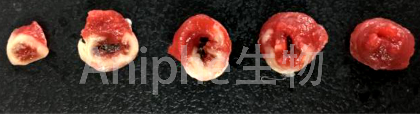
Image source: Aniphe BioLab
Evans Blue + TTC double staining
Evans Blue staining at a glance; image source: Aniphe BioLab.
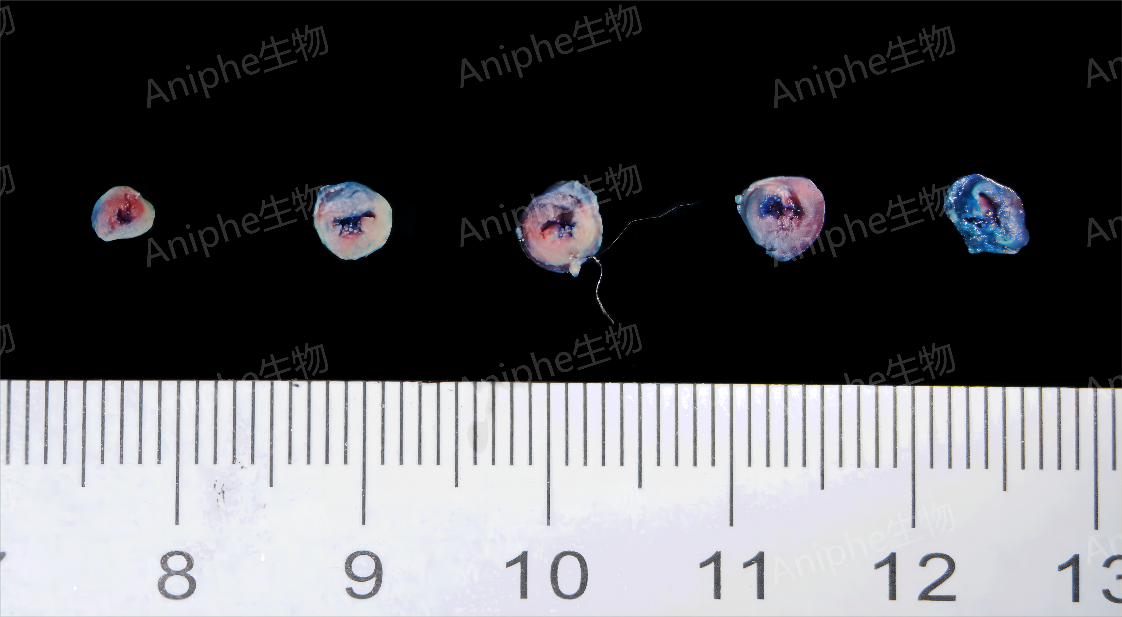
Image source: Aniphe BioLab
Using Evans Blue plus TTC double staining, the non‑ischemic region in the model group appears blue, the infarcted area is white, and the at‑risk zone remains unstained, retaining the native red color of myocardial tissue.
The Evans Blue + TTC double-staining method is a combined technique that can Simultaneously assess myocardial cell integrity and metabolic activity. , primarily used for animal assessment In a cardiac ischemia-reperfusion injury model, the relative sizes of the at-risk zone and the infarcted area allow for visualization of the extent of myocardial damage. , thereby enabling the localization and analysis of myocardial infarction–related areas.
Pathological staining
In the control group, the tissue architecture was normal, with no obvious inflammatory cell infiltration. In the model group, blue collagen deposition was observed at the infarct site, indicating marked fibrosis.
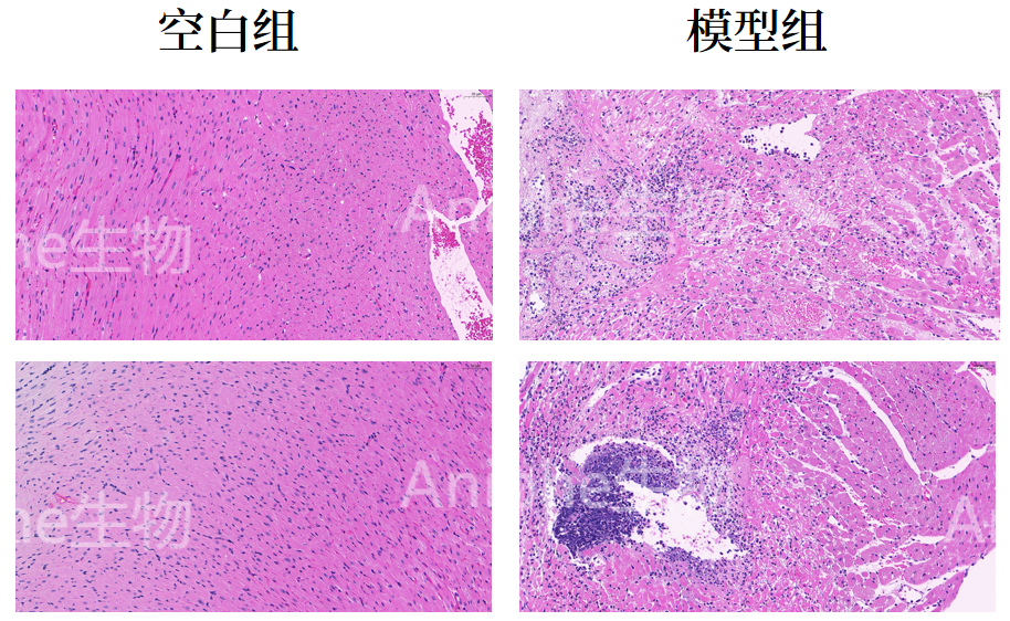
Image source: Aniphe BioLab
Precautions
The construction of a myocardial ischemia (MI) model in rats is similar to that in mice; an appropriately sized suture should be selected based on the rat’s body weight. In addition, the following details should be carefully observed during the procedure:
01 Complete coronary artery ligation and close the chest wall as soon as possible to reduce the risk of life‑threatening complications—such as arrhythmias and pneumothorax—that may arise from prolonged thoracic exposure.
02 The coronary arteries are small, with a rich network of collateral circulation that is difficult to visualize; therefore, the experimenter must perform the procedure with precision, ensuring sufficient myocardial infarction while avoiding an excessively large infarct size that could lead to mortality. It is advisable to ligate at a consistent level to establish a more uniform model.
03 The ventricular wall is relatively thin, so the operator must carefully control the puncture depth to prevent excessive penetration that could directly perforate the ventricular wall and lead to massive hemorrhage.
——————————————
Myocardial ischemia-reperfusion injury (MIRI) refers to the phenomenon in which myocardial cells suffer ischemic damage due to various causes, and upon restoration of blood supply, the ultrastructural, functional, metabolic, and physiological impairments of the myocardium are further exacerbated.
Compared with the model of chronic myocardial ischemia, in the latter, a 7-0 suture is used to tie a tight knot to permanently ligate the left anterior descending artery (LAD), thereby inducing permanent myocardial infarction. In contrast, the myocardial ischemia–reperfusion injury model employs a 7-0 suture tied in a loose knot or a ligature rod for ligation; after a specified period of ischemia, the loose knot is released and the ligature rod is removed to restore blood flow, thereby reperfusing the previously ischemic myocardial tissue.
References:
[1] Ding Yiming, Gao Jie, Xing Yuanming, et al. Establishment and systematic evaluation of a mouse model of myocardial infarction [J]. Chinese Journal of Laboratory Animal Science, 2022, 30(07):873-879.
Jiangsu Aniphe BioLab Inc., established in 2018, is committed to becoming an internationally renowned preclinical pharmacodynamic evaluation company in the new era of biopharmaceutical development. Aniphe BioLab has built a comprehensive SPF‑grade laboratory animal center that integrates animal breeding and husbandry, disease model generation, and in vivo experimentation, covering areas such as oncology, neuroscience, and metabolism. The center houses over 5,000 cages for mice and rats. With a highly skilled technical team and abundant animal resources, Aniphe BioLab offers a wide array of disease‑specific animal models, including mouse and rat models of cerebral ischemia–reperfusion (MCAO/R), myocardial infarction/myocardial ischemia (MI), thoracic aortic constriction (TAC), human‑derived tissue xenograft tumor models (PDX), cell‑line xenograft tumor models (CDX), spinal cord injury (SCI), epilepsy, and Alzheimer’s disease (AD). In addition, the company provides pharmacological and efficacy studies tailored to these disease models. We warmly welcome clients to contact us for more information.
Aniphe BioLab Inc.
Address:
Runcheng, No. 8 Longtai Road, Jiangbei New Area, Nanjing City, Jiangsu Province
Building 8, Science Park
Postal Code: 210007
Phone:025-8520 8061
Email:info@aniphebio.com(Consultation)
Email:order@aniphebio.com(Order)


WeChat Official Account
Video Account
 Su Public Security Bureau Internet Information Service Filing No. 32010402001421