
-
Research Services<a class="dhimg1" href="/shichangdetail1/9.html" target="_self" rel="nofollow"> <img src="https://omo-oss-image.thefastimg.com/portal-saas/pg2026042316544414203/cms/image/8926ff01-64bc-4928-a995-396bdb9d8102.jpg" alt="Aniphe BioLab"> </a> <a class="dhimg2" href="/shichangdetail/8.html" target="_self" rel="nofollow"> <img src="https://omo-oss-image.thefastimg.com/portal-saas/pg2026042316544414203/cms/image/26342b34-4f1e-4d28-9166-7b685e6d70c3.jpg" alt="Aniphe BioLab"> </a>


-
Preclinical CRO<a class="dhimg1" href="/shichangdetail/6.html" target="_self" rel="nofollow"> <img src="https://omo-oss-image.thefastimg.com/portal-saas/pg2026042316544414203/cms/image/cc43bac1-07d9-4340-9677-fbe01c51330a.jpg" alt="Aniphe BioLab"> </a> <a class="dhimg2" href="/shichangdetail/10.html" rel="nofollow" target="_self"> <img src="https://omo-oss-image.thefastimg.com/portal-saas/pg2026042316544414203/cms/image/f1d8f739-0d36-4cb8-adeb-26de791d909c.jpg" alt="Aniphe BioLab"> </a>


-
-
-
-


Latest News
Focusing on cutting-edge technologies and research advancements,
sharing Aniphe BioLab’s achievements in scientific innovation,
Professional insights
and practical experience in the fields of laboratory animals and preclinical research.
Publish an article
21-Day Rapid Modeling: Construction of an Orthotopic VX2 Rabbit Hepatocellular Carcinoma Model—Comprehensive Analysis of Pathological Findings, Tumor Growth Curves, and Imaging Features
Primary liver cancer (PLC) is one of the most common malignancies. The VX2 rabbit orthotopic liver cancer model exhibits tumor progression that closely resembles human hepatocellular carcinoma, making it an ideal experimental model for liver cancer research. Therapeutics and drugs developed in this model are of great significance for guiding clinical decision‑making in selecting appropriate treatment strategies.
The VX2 tumor was initially derived from rabbit squamous cell carcinoma induced by the Shope papillomavirus through repeated passages. It exhibits strong invasiveness and rapid proliferation, and can spontaneously metastasize via the portal vein or hepatic artery to the lungs, lymph nodes, and other sites, making it one of the few animal models capable of mimicking the metastatic behavior of hepatocellular carcinoma.
Construction of an Orthotopic VX2 Rabbit Hepatocellular Carcinoma Model
| Open-Access Implantation Method
① Laboratory animals
New Zealand White rabbits, weighing 2.5–3.5 kg and aged 5–6 months, have a moderate body size and excellent tolerance, making them well suited for surgical procedures.
② Experimental Methods
Experimental rabbits were fasted for 12 hours prior to inoculation, with free access to water.
Anesthesia was administered, and an abdominal incision was made to expose the left lobe of the liver. The tumor mass was injected into the left lobe via a puncture needle; after packing with gelatin sponge, hemostasis was achieved by applying pressure with sterile cotton swabs.

Reinsert the liver into the body and close the incision. Administer a prophylactic antibiotic (e.g., enrofloxacin) for 3 days postoperatively.
④ Precautions
This method features a short tumor‑formation time, a high implantation success rate, localized tumor growth, and minimal ectopic implantation.
| Experimental Results
① Pathological tissue
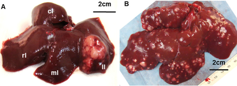
② Growth curve:
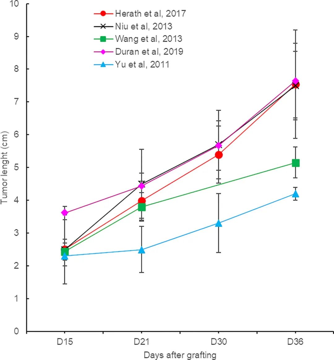
② HE results:
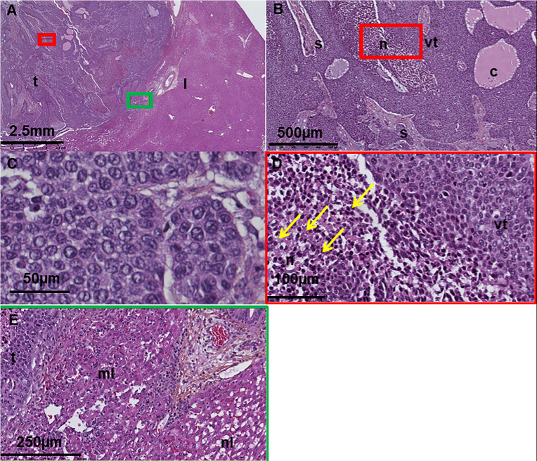
Digital histological sections of VX2 tumors in the liver at different magnifications; image source [1].
③ Imaging features
CT :
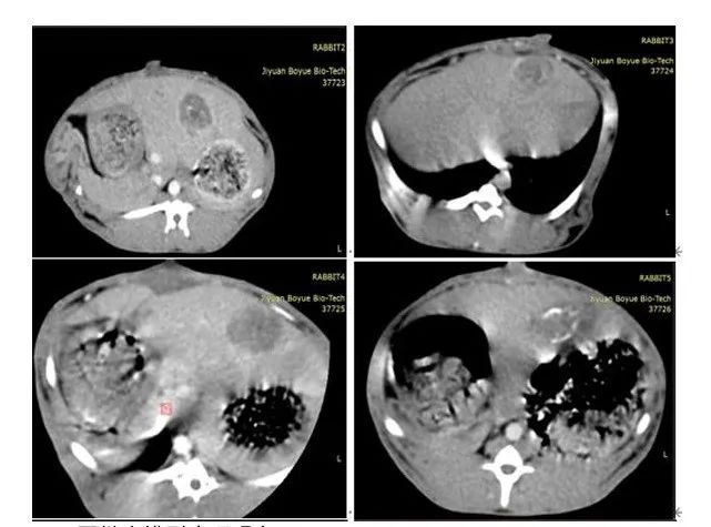
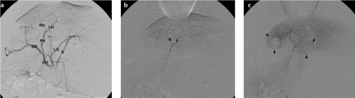
Image source [1]
Procedure:
The procedure is minimally invasive and carries no risk of peritoneal implantation; the tumor models are typically solitary nodules, facilitating imaging‑based monitoring. The success rate exceeds 95%, comparable to that of the open‑abdominal approach. Larger rabbits should be selected to facilitate the procedure, and puncture must be precisely directed to avoid the interlobar spaces, thereby preventing inadvertent tumor implantation.
| Tumor cell suspension injection method
Operation Steps:
Model features:
Summary
Aniphe BioLab Inc.
Address:
Runcheng, No. 8 Longtai Road, Jiangbei New Area, Nanjing City, Jiangsu Province
Building 8, Science Park
Postal Code: 210007
Phone:025-8520 8061
Email:info@aniphebio.com(Consultation)
Email:order@aniphebio.com(Order)


WeChat Official Account
Video Account
 Su Public Security Bureau Internet Information Service Filing No. 32010402001421