
-
Research Services<a class="dhimg1" href="/shichangdetail1/9.html" target="_self" rel="nofollow"> <img src="https://omo-oss-image.thefastimg.com/portal-saas/pg2026042316544414203/cms/image/8926ff01-64bc-4928-a995-396bdb9d8102.jpg" alt="Aniphe BioLab"> </a> <a class="dhimg2" href="/shichangdetail/8.html" target="_self" rel="nofollow"> <img src="https://omo-oss-image.thefastimg.com/portal-saas/pg2026042316544414203/cms/image/26342b34-4f1e-4d28-9166-7b685e6d70c3.jpg" alt="Aniphe BioLab"> </a>


-
Preclinical CRO<a class="dhimg1" href="/shichangdetail/6.html" target="_self" rel="nofollow"> <img src="https://omo-oss-image.thefastimg.com/portal-saas/pg2026042316544414203/cms/image/cc43bac1-07d9-4340-9677-fbe01c51330a.jpg" alt="Aniphe BioLab"> </a> <a class="dhimg2" href="/shichangdetail/10.html" rel="nofollow" target="_self"> <img src="https://omo-oss-image.thefastimg.com/portal-saas/pg2026042316544414203/cms/image/f1d8f739-0d36-4cb8-adeb-26de791d909c.jpg" alt="Aniphe BioLab"> </a>


-
-
-
-


Latest News
Focusing on cutting-edge technologies and research advancements,
sharing Aniphe BioLab’s achievements in scientific innovation,
Professional insights
and practical experience in the fields of laboratory animals and preclinical research.
Publish an article
Methods for establishing a pulmonary fibrosis (PF) model—understand mouse CT imaging changes at a glance!
Common animal models of pulmonary fibrosis
At present, many inducers are used in model making, such as Bleomycin, paraquat, high-concentration oxygen, asbestos, and ionizing radiation, among others. All can cause lung injury, ultimately leading to pulmonary fibrosis.
Among them Bleomycin is the most commonly used. Bleomycin is an aminoglycopeptide antibiotic isolated from the culture broth of Streptomyces verticilli, exhibiting potent antitumor activity and serving as a first-line anticancer agent. Its adverse effects can lead to pulmonary fibrosis and associated clinical manifestations.

Protocol for Establishing a Mouse Model of Pulmonary Fibrosis
Aniphe BioLab Bleomycin-Induced Case Presentation:
| Laboratory Animals
C57 male, 10–12 weeks old.
| Modeling Method
| Experimental Evaluation System
① Lung coefficient:
The lung mass was weighed, and the lung coefficient was calculated as follows: Lung coefficient (%) = Lung mass (g) / Body mass (g) × 100%.
② Lung HE staining :
Following the experiment, mouse lung tissues were collected and fixed for hematoxylin–eosin staining.
| Experimental Evaluation System
① Pathological tissue :

Pathological tissue; image courtesy of Aniphe BioLab.
② CT findings Result display :

CT results; image source: Aniphe BioLab.
② HE results show:
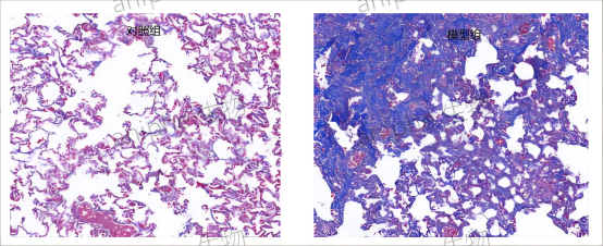
HE results; image source: Aniphe BioLab.
| Model Features:
Intratracheal administration of bleomycin can directly injure alveolar epithelial cells, and a single dose is sufficient to induce fibrosis within a short time.
| Other induction methods
① Bleomycin-induced pulmonary fibrosis model
Studies have shown that intraperitoneal injection of 20 mg/kg in rats or 10 mg/kg in mice can induce the model after a certain period (onset within 5–9 days, peaking at 2–3 weeks). Alternatively, a high oral dose of bleomycin can be used to establish the model: administration of 50 mg/kg by gavage leads to pulmonary fibrosis within 120 hours.
The pathogenic mechanism involves the substance’s absorption into the body, where it undergoes complex biochemical processes that generate large amounts of oxygen free radicals, leading to lipid peroxidation in organ and tissue cells and subsequently inducing tissue fibrosis. Alveolar cells exhibit a strong active uptake and accumulation of paraquat; consequently, the lungs suffer the most severe damage and are particularly prone to developing pulmonary fibrosis.
② Amiodarone-induced pulmonary fibrosis model
Amiodarone (AD) is a highly effective antiarrhythmic agent that is associated with a variety of adverse effects. Studies have shown that AD‑induced pulmonary toxicity primarily manifests as the accumulation of foamy cells within the alveolar spaces, hyperplasia of type II pneumocytes, and pulmonary interstitial fibrosis.
One study established a rat model of amiodarone‑induced pulmonary toxicity by administering 0.1 mL of amiodarone hydrochloride (1.25 mg) in saline via intratracheal instillation once daily for 5 consecutive days. On day 28, marked proliferation of fibroblasts was observed around blood vessels and bronchi, accompanied by a significant increase in collagen deposition; collagen fibers within the adjacent lung parenchyma were also markedly augmented, and numerous foam cells were found to be aggregated. Alveolar septal thickening was evident, type II pneumocytes exhibited prominent hyperplasia, and pulmonary fibrosis had developed.
③ Oleic acid–induced pulmonary fibrosis model
The mechanisms underlying oleic acid–induced pulmonary fibrosis are primarily as follows: under the influence of inflammatory mediators and chemokines, neutrophils are activated to adhere to endothelial surfaces, migrate toward the site of inflammation, and engage in phagocytosis, degranulation, and the respiratory burst, thereby releasing lysosomal enzymes, reactive oxygen species, and other bioactive metabolites. While these processes eliminate pathogenic stimuli, they also inflict tissue damage. Concurrently, elevated levels of malondialdehyde (MDA) promote protein cross‑linking and denaturation, induce DNA fragmentation, and disrupt the structural integrity of lung tissue, driving the progressive proliferation of fibrous tissue and ultimately precipitating pulmonary fibrosis.
④ Silica-induced pulmonary fibrosis model
Silica dust is an industrial particulate that, upon long-term inhalation, can cause severe pulmonary damage. At present, the use of silica to establish pulmonary injury models is primarily focused on replicating silicosis. Typically, such models are generated by directly instilling a silica suspension into the trachea of experimental animals.
⑤ Asbestos-induced pulmonary fibrosis model
Asbestos particles are a workplace environmental toxin that, upon inhalation into the alveoli, induce localized alveolar inflammation, release cytokines, and stimulate fibroblast proliferation and the excessive deposition of extracellular matrix. Repeated exposure ultimately triggers pulmonary fibrosis. In one study, 1 mL of an asbestos–saline suspension at a concentration of 20 g·L⁻¹ was directly instilled into the lungs of rats via a laryngoscope inserted into the trachea, followed by gentle massage of both lungs for a brief period, successfully establishing a rat model of pulmonary fibrosis.
⑤ Radiation‑induced pulmonary fibrosis model
In the course of scientific research on how to prevent and treat radiation‑induced lung injury, it is essential to establish relevant animal models of radiation‑induced pulmonary fibrosis. One study utilized a SIEMENS Primus Hi high‑energy linear accelerator to deliver a single 12 Gy dose to the lungs of mice via the anterior thoracic field. The results showed that, by Masson’s trichrome staining, collagen fiber deposition began to appear in the lung tissue of irradiated mice approximately four weeks after exposure, and at 16 and 24 weeks, distinct focal areas of fibrosis were readily visible under light microscopy, indicating successful establishment of the pulmonary fibrosis model.
Summary
The methods for establishing animal models of pulmonary fibrosis have become increasingly diverse, and the associated techniques have grown ever more sophisticated. Selecting an appropriate model is of great significance for investigating the pathological processes and underlying mechanisms of pulmonary fibrosis.
Aniphe BioLab Inc.
Address:
Runcheng, No. 8 Longtai Road, Jiangbei New Area, Nanjing City, Jiangsu Province
Building 8, Science Park
Postal Code: 210007
Phone:025-8520 8061
Email:info@aniphebio.com(Consultation)
Email:order@aniphebio.com(Order)


WeChat Official Account
Video Account
 Su Public Security Bureau Internet Information Service Filing No. 32010402001421